CLINICAL RESEARCH
Clinical and Radiographic Evaluation of the OMICRON 2® Total Knee Prosthesis: A Multicenter Study With Long-Term
Follow-up
Carlos Vega,*,**
Favio Moruno, *,** Javier Megey, * Wilmer Jimenez, * Carlos F. Satragno#
*Knee Unit,
Hospital Municipal Central
de San Isidro “Dr. Melchor
Á. Posse“, Buenos
Aires, Argentina
**Hip and Knee Unit,
Hospital Zonal de Agudos “Dr. Carlos A. Bocalandro“, Buenos Aires, Argentina
#Hip and Knee Unit, Hospital Municipal
“Bernardo Houssay”, Buenos
Aires, Argentina
ABSTRACT
Introduction: Total knee
arthroplasty is the ideal treatment for severe osteoarthritis. The OMICRON 2® system, introduced in
Argentina in 2009, incorporated improvements in implant design and
instrumentation to optimize and prolong implant survival. Materials and Methods: A
retrospective, longitudinal, observational study included 1,346 total knee
arthroplasties performed between 2009 and 2024 at three hospitals
in the province of Buenos Aires. Complete
clinical and radiographic follow-up data are presented for 616 patients. The
Knee Society Score (KSS) and Western
Ontario and McMaster Universities Osteoarthritis Index (WOMAC) were used to assess function
over time, together
with standardized radiographs. Implant survival was defined as retention
of the implant without revision and was assessed using Kaplan-Meier curves. Results: The
mean age was 71.6 years. The most common
diagnosis was primary
knee osteoarthritis (86%).
Implant survival at 15 years was 92.4%. The KSS improved
from
37.5 preoperatively to 85.1 at 15
years, and the WOMAC score improved
from 89.2 to 15.5 over the same period. At
the end of follow-up, 89.8% of patients had no signs of loosening, and only 2%
showed minimal femoral changes. Complications
included infections (2.7%), septic
loosening, periprosthetic fractures, and instability. Conclusion: The OMICRON
2® system demonstrated good clinical and radiographic performance, with high long-term implant
survival, and is a reliable
option for total
knee arthroplasty. Keywords: Knee
arthroplasty; osteoarthritis; OMICRON 2®; survival.
Level of Evidence: IV
Análisis clínico-radiológico de la prótesis total de rodilla
OMICRON 2®. Estudio multicéntrico con seguimiento a largo plazo.
RESUMEN
Introducción: La artroplastia total
de rodilla es el tratamiento ideal para la artrosis severa.
El sistema OMICRON
2®, introducido en la Argentina, en 2009, desarrolló mejoras en el diseño e instrumental destinadas a optimizar y aumentar la supervivencia del implante. Materiales y Métodos: Se llevó a cabo un estudio
observacional, longitudinal y retrospectivo sobre 1346 artroplastias totales de rodilla
realizadas entre 2009 y 2024, en tres hospitales de la provincia de Buenos Aires. Presentamos
el seguimiento clínico-radiológico completo de 616 pacientes. Se utilizaron los cuestionarios KSS y WOMAC
para valorar la funcionalidad en el
tiempo con radiografías estandarizadas. La
supervivencia se definió como la permanencia del implante en el sitio, sin
revisión, evaluado mediante curvas
de Kaplan-Meier. Resultados: La
media de edad era de 71.6 años. El diagnóstico más frecuente fue gonartrosis primaria (86%). La supervivencia del implante a los 15
años fue del 92,4%. El puntaje KSS
mejoró de 37,5 antes de la cirugía a 85,1 a los 15 años,
y el puntaje WOMAC, de 89,2 a 15,5. El 89,8% no tenía signos
de aflojamiento al final del seguimiento; solo un 2% presentó cambios mínimos
femorales. Las complicaciones fueron
infecciones (2,7%), aflojamiento séptico, fracturas periprotésicas e inestabilidad. Conclusión: El sistema
OMICRON 2® tuvo un buen rendimiento clínico
y radiológico, con una alta supervivencia a largo plazo; es una opción confiable en la artroplastia
total de rodilla.
Palabras clave: Artroplastia de rodilla; artrosis; OMICRON 2®; supervivencia.
Nivel de Evidencia: IV
INTRODUCTION
Total knee arthroplasty (TKA)
is currently the gold standard
for treating severe
osteoarthritis.1,2 As the procedure has become increasingly common over the past 30 years, implants
and instrumentation have improved significantly.3 Outcomes are assessed using
several variables, including implant survival, kinematics, function, and
patient satisfaction.4-6
The
profile of patients considered candidates for TKA varies widely across
publications in terms of age and disease progression, with increasingly younger
patients undergoing the procedure.7,8
For
patients with osteoarthritis who have a life expectancy of 20–40 years,
reliable long-term follow-up data on
TKA provide a sound basis for decision-making. Long-term follow-up studies of
TKA performed within the past decade therefore warrant greater attention,
despite the logistical challenges and limitations.9,10 Evidence on implants with an established record
of long-term use can also help assess the quality and performance of new devices.11
Implant
designers and surgeons continue to develop new implants and surgical techniques
that may increase implant survival and improve outcomes after TKA.
The
OMICRON 2® TKA system, manufactured by Ortopedia IMECO S.A., was introduced to
the Argentine market in 2009 with
certification from the National Administration of Drugs, Food and Medical
Devices (AN-MAT). It has since been distributed nationwide.
This study aimed to evaluate the long-term clinical
and radiographic outcomes
and implant survival
rates of the OMICRON 2® knee prosthesis.
MATERIALS AND METHODS
Implant
The
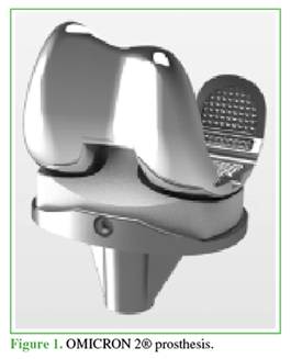
implant has a posterior-stabilized design with an integrated post that
compensates for the absence of the posterior
cruciate ligament (Figure 1).
The
design was refined from its predecessor, the OMICRON 1® prosthesis, to achieve
mechanical alignment while balancing bone resection (bone balancing) with the
balancing of ligamentous and capsular structures (gap balancing). It features
more anatomically shaped components designed for cemented fixation.
The femoral
components are made of a cobalt-chromium-molybdenum alloy
and have a single radius
of rotation to improve
tibiofemoral joint congruity. The femoral component was modified to provide a more anatomical shape and trochlear orientation, with distinct right and left
components. The trochlear groove was deepened to allow smoother patellar
tracking. A smaller intercondylar
box, proportional to the size of the femoral component, helps preserve bone
stock.
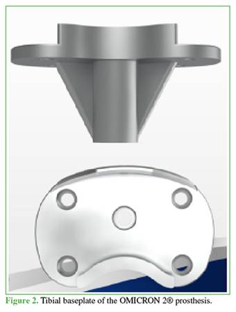
The
tibial components are also made of a cobalt-chromium-molybdenum
alloy and are modular, with a short central stem.
The
tibial baseplate is bilaterally symmetrical, with curved posterior edges
that provide an appropriate
fit to the tibia (Figure 2).
The keel has two antirotation fins and is smaller
than that of the previous
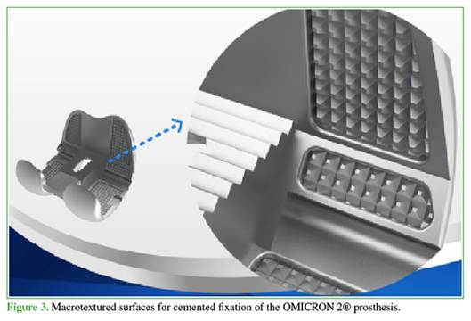
implant. The inner surface
of the femoral component and the undersurface of the tibial component
are macrotextured to improve
cement fixation (Figure 3).
The
polyethylene is highly cross-linked to prevent early wear and incorporates 3°
of posterior slope. Femoral alignment was established using an intramedullary
guide, and tibial alignment using an extramedullary guide. The posterior slope
could be increased to 7° according to the patient’s characteristics and the
surgeon’s judgment.
The
instrumentation is based on an anatomical technique guided by mechanical
alignment, allowing sequential soft tissue release.
Study Implementation
Implant
survival depends not only on implant design but also on several factors related
to surgical technique. Hospitals in the province of Buenos Aires were therefore
considered using uniform selection criteria.
During
much of the study period, the OMICRON 2® prosthesis was supplied to 16 public
hospitals in the province of Buenos Aires through the Comprehensive Medical
Care Program (PAMI), allowing large-volume distribution. Hospitals from this
group were selected according to the following criteria:
• Availability of clinical
and radiographic records throughout follow-up.
• Use of the same surgical
approach, implant, and instrumentation.
• Use of the same measured
resection technique with balancing of the flexion and extension
gaps in all cases.
• A standardized cementing
technique aimed at optimizing preparation of the bone bed.
• Use of the transepicondylar axis to determine
external rotation of the femoral component in all procedures.
• Tibial component rotation guided by the tibial tubercle.
• Use of the same brand of domestically manufactured bone cement.
Based
on these criteria, a retrospective longitudinal observational study was
conducted at three hospitals in the province of Buenos Aires: Hospital Municipal Central de San Isidro “Dr. Melchor Á. Posse,” Hospital Zonal de Agudos “Dr.
Carlos A. Bocalandro,” and Hospital Municipal “Bernardo Houssay.”
A
total of 1,346 TKAs were performed between 2009 and 2024. Only 616 patients
completed the clinical and radiographic assessment; the remaining patients were
lost to follow-up or had died. Patients were classified as lost to follow-up
when they missed follow-up visits and could not subsequently be contacted.
Deaths that could be confirmed were documented through hospital records and
telephone contact with relatives or acquaintances.
Patients who underwent primary
TKA were included, and revision procedures were excluded. Implant
survival was defined as retention of the implant in situ at the final
follow-up visit.
Statistical Methods
Clinical
and functional outcomes were recorded in a standardized manner at each hospital
by multiple examiners who were blinded during the study. Assessments used questionnaires specifically intended for these
patients, administered before and after surgery.
The database was combined with a retrospective review of medical
records to collect demographic data (age, sex, and body mass index) and
surgical data.
A
Kaplan–Meier curve was used to determine implant survival at 15 years after
implantation. Patient function was assessed using the Knee Society Score (KSS)
and the Western Ontario and McMaster
Universities Osteoarthritis Index (WOMAC).
Permission to obtain patient data was requested from the ethics committee at each hospital.
In
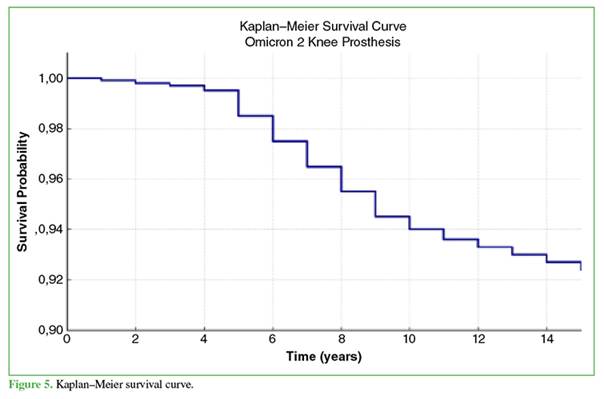
terms of implant survival, the success rate was 92.4% (p = 0.924) among the 616
patients followed in this study. The minimum required sample size was n = 300
patients, yielding a margin of error of 0.03%, a standard level of precision
for clinical studies.
Patient Population
The
mean age at implantation was 71.6 years; 67.2% of patients were women and 32.8%
were men. Mean weight was 80.9 kg, and mean body mass index was 29.2 kg/m2 (Table
1).
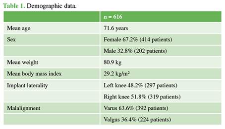
Surgery was performed on the left knee in 48.2% of cases and the right
knee in 51.8%.
Varus was the most common malalignment (71.1%), followed by
valgus (28.9%). All patients had
competent ligaments, allowing the use of primary knee prostheses.
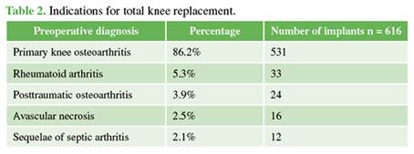
The most common diagnosis leading to surgery
was primary knee osteoarthritis (86%),
followed by rheumatoid arthritis (6%),
posttraumatic osteoarthritis (4%),
sequelae of femoral
osteonecrosis (2.5%), and osteoarthritis secondary to septic arthritis (2%) (Table 2).
The
730 patients without complete follow-up were slightly older than those who
completed follow-up, with a mean age of 73.8 years. There were no statistically significant differences in sex
distribution or body mass index (p > 0.05).
Surgical Technique
Preoperative
planning included weight-bearing anteroposterior radiographs of both knees, a
lateral view at 30° of flexion, and an axial patellar view to provide a
comprehensive assessment of the affected joint.12
Standard prophylaxis included antibiotics 30 minutes before
skin incision and antithrombotic prophylaxis in the immediate
postoperative period.
All
procedures were performed through a medial parapatellar approach.
The prostheses were implanted using a measured
resection technique and standard instrumentation. The patella was everted and resurfaced at the surgeon’s
discretion, depending on the characteristics of the bone. As a general rule,
resurfacing was performed in cases of extensive patellar cartilage destruction.
It was contraindicated when patellar thickness was <20 mm because of the
risk of fracture.13 The patella was resurfaced in 61.5% of patients.
All
components were cemented, with particular attention to preparing a clean, dry
cancellous bone bed to improve cement interdigitation. Cement was applied to
both the bone surface and the implant, including the keel, which was cemented
in all cases.
RESULTS
Two independent surgeons at each hospital performed
the radiographic assessments. Implant survival and stability
were assessed by examining standardized zones around the components for signs
of loosening.
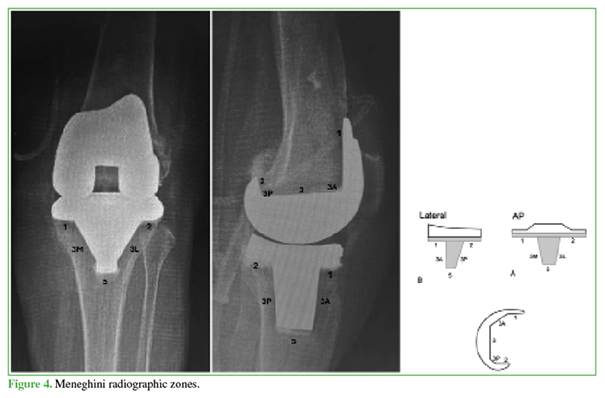
The system described by Meneghini et al. and Kumar et al. was used to assess implant
stability radiographically (Figure 4).9,14
Radiographic
findings at the final follow-up were retrieved from each hospital’s digital
database for the 616 patients in the study population. Radiographs for the
remaining patients were unavailable because of loss to follow-up. Deaths during
follow-up were identified through hospital records and confirmed by telephone
contact with relatives.
At the final follow-up, 89.8% of implants
showed no radiographic changes or signs of loosening. Changes suggestive of osteolysis measuring <1 mm were observed in
zones 1 and 2 around the tibial component in 8.2% of implants. The remaining 2%
showed minimal changes measuring <2 mm in zone 1 around the femoral
component, without clear signs of loosening.
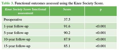
Functional
outcomes were assessed using the KSS (Table 3).
The total score (maximum, 100) improved significantly from a preoperative mean
of 37.5 to 91.6 at 1 year (p < 0.001), 90.2 at 5
years (p < 0.001), 87.9 at 10 years (p < 0.001), and 85.1 at 15 years. Our findings are consistent with those reported
in the current literature. The WOMAC score (worst possible score, 96) also
improved, from a preoperative mean of 89.2 to 15.5 at the final follow-up.
A key
strength of our findings on KSS and WOMAC score improvements is that they are
based on long-term clinical and radiographic follow-up.
The
main limitation was the progressive loss of patients to follow-up. Although substantial, this loss is
unsurprising and consistent with that reported
in other long-term
studies. Nevertheless, the 95% confidence interval for implant
survival at 15 years was 90.31%–94.49%, indicating high statistical precision,
with a margin of error of 2.09%, and supporting the robustness of the study.
The
design features of the OMICRON 2® total knee replacement system have remained
unchanged over the years. Its favorable functional outcomes and 15-year implant
survival reflect the success of its design. The
OMICRON 2® prosthesis therefore serves as the benchmark for comparing TKA
systems at the participating institutions.
Complications
Complications are detailed in Table 4, although
most were unrelated to the implants.
Twenty-three revision
procedures were performed. Five were for septic loosening
and were managed
with two-stage revision.
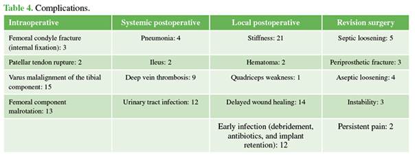
Three periprosthetic fractures involved substantial loss of bone stock and required revision
to a more constrained prosthesis.
Aseptic loosening
led to revision in 10 patients. The tibial component
had been placed in varus in seven patients,
and the femoral component was malrotated in three, resulting in osteolysis and
early loosening.
Three
patients required further surgery for instability and two for persistent
anterior knee pain. Neither of the patients with anterior knee pain had
undergone patellar resurfacing.
The infection
rate was 2.7%,
with chronic infections in five patients
and acute infections in 12. The acute infections resolved
with early debridement, antibiotics, and implant retention.
DISCUSSION
This
study reports the long-term outcomes of a consecutive series of 616 patients
treated between 2009 and 2024.
Overall, the 15-year implant survival rate of 92.4% is favorable (Figure 5).
Plain
radiographs are the most widely used imaging modality for assessing component
alignment, although three-dimensional computed tomography is the reference
standard.15,16 We used the Knee Society TKA roent-genographic
evaluation and scoring system,17 an accepted tool for radiographic assessment.18,19
We believe
that restoring alignment plays an important role in improving implant survival 20 and functional outcomes.21,22 Patient age may also influence implant survival. Some series of cemented knee replacements in young
patients with rheumatoid arthritis have reported excellent long-term survival
rates.23,24 However, these favorable results may be
influenced by the lower activity levels of patients with inflammatory
arthritis.
Younger
patients generally have greater physical demands and a longer life expectancy,
placing greater mechanical stress on the implants.
Roberts
et al. found that implant survival was significantly higher in women and older
patients.25 Julin et al. reported 5-year survival rates of
92% and 95% in patients aged <55 years and 56–65 years, respectively,
compared with 97% in those aged >65 years.26 Younger
patients had higher revision rates for noninfectious reasons. Keenan et al.
reported 10-year survival after cemented TKA
in patients aged <55 years at surgery and compared their functional outcomes
with those of patients aged >55 years.27
The
original cohort of 1,346 patients decreased over the study period. By 12–15
years, 106 patients had died and 624 had been lost to follow-up. Those lost to
follow-up were older than 85 years at the time of assessment, so it is reasonable to assume that many
had died.
Declining
patient numbers are common in long-term joint replacement studies, reducing the
usefulness of functional scores as a discriminating measure.28,29 Some
studies report that 82% of TKAs last approximately 25 years.30 Callaghan
et al. reported on the 26 surviving patients from an initial cohort of 86.
Despite an excellent KSS of 89, the function score had declined to 67.31 Reports with a
minimum follow-up of 15 years
after TKA are scarce, and several
concern implants that are no longer commonly used. Van Loon et al. followed 102 cemented kinematic TKAs for 10–15
years and reported a survival rate of 62% at 14 years. The main reasons for
revision were deep infection
and wear.32 Buechel reported
a survival rate of 83% at 16 years.GG Ito et al. examined 36 knees
in 25 patients with rheumatoid arthritis and reported a 15-year survival rate
of 93.7%.34
Our
results are therefore comparable with those reported in the international
literature. However, we could not compare them with findings from Argentina, as
no long-term studies of domestically manufactured prosthetic implants have been
reported.
This
study has several strengths and limitations that should be considered when
interpreting its findings. A major strength is the length of follow-up, which
allows assessment of the implant’s long-term durability, with implant survival
serving as a key indicator of clinical success. The observational design provides information on how the
prosthesis performs under routine clinical conditions, encompassing a diverse
population and reflecting everyday surgical practice. Finally, the sample size
and availability of longitudinal data allow reliable statistical analyses.
Nevertheless,
the study has limitations inherent to its design. The main limitations are the absence of a control group and the
high rate of loss to follow-up, which may introduce bias, particularly
selection and information bias. Reliance on retrospective clinical records also
poses a risk of bias because these records may contain inaccurate data.
Changes
or errors in the surgical techniques used during the study period may also have
influenced the outcomes. For example, the five cases of aseptic loosening were
associated with surgical errors involving incorrect component orientation.
These
factors should be considered when assessing the validity and applicability of
the findings. Although the
retrospective design provides valuable insight into the prosthesis’s
performance in routine clinical practice, studies with more controlled designs
are needed to support its conclusions and confirm the findings.
CONCLUSIONS
We report
a favorable implant
survival rate of 92.4% in a series
of 616 TKAs using the OMICRON 2® system
after 15 years of follow-up.
The
system’s distinctive design features have remained unchanged throughout its
relatively long clinical history. In other systems, these features are often
modified before long-term results are published.
The
OMICRON 2® system continues to show favorable long-term implant survival at a
mean follow-up of 15 years.
REFERENCES
1. Jauregui JJ, Cherian JJ, Pierce TP, Beaver WB, Issa K, Mont MA. Long-term survivorship and clinical outcomes following total knee arthroplasty. J Arthroplasty 2015;30(12):2164-6. https://doi.org/10.1016/j.arth.2015.05.052
2. Argenson JN, Boisgard S, Parratte S, Descamps S, Bercovy M, Bonnevialle P, et al. Survival analysis
of total knee arthroplasty at a minimum
10 years follow-up: A multicenter French
nationwide study including 846 cases. Orthop
Traumatol Surg Res 2013;99(4):385-90. https://doi.org/10.1016/j.otsr.2013.03.014
3. Parratte S, Pagnano MW, Trousdale RT, Berry DJ. Effect of postoperative mechanical axis alignment on the fifteen-year survival of modern, cemented
total knee replacements. J Bone Joint
Surg Am 2010;92(12):2143-9. https://doi.org/10.2106/JBJS.I.01398
4. Aujla
RS, Esler C. Total knee arthroplasty for osteoarthritis in patients <55 years of age: A systematic review,
J Arthroplasty 2017;32(8):2598-2603.e1. https://doi.org/10.1016/j.arth.2017.02.069
5. Longo
UG, Ciuffreda M, Mannering N, D’Andrea V, Locher J, Salvatore G, et al. Outcomes of posterior-stabilized
compared with cruciate-retaining total knee arthroplasty J Knee Surg 2018;31(04):321-40. https://doi.org/10.1055/s-0037-1603902
6. Benazzo F, Ghiara M, Rossi SMP, Pruneri E, Tiwari T, Perelli S. Clinical and radiological analysis
of a personalized total knee arthroplasty system design. Int Orthop 2019;43(5):1113-21. https://doi.org/10.1007/s00264-018-4095-4
7. Callaghan JJ, Martin CT, Gao Y, Andrew J, Pugely AJ, Liu SS, et al. What can be learned
from minimum 20-year followup
studies of knee arthroplasty? Clin Orthop
Relat Res 2015;473(1):94-100. https://doi.org/10.1007/s11999-014-3744-1
8. Becker
R, Doring C, Denecke A, Brosz M. Expectation, satisfaction and clinical outcome
of patients after
total knee arthroplasty Knee Surg Sports Traumatol
Arthrosc 2011;19(9):1433-41. https://doi.org/10.1007/s00167-011-1621-y
9. Meneghini RM, Mont MA, Backstein DB, Bourne RB, Dennis DA, Scuderi GR. Development of a modern knee Society Radiographic Evaluation System and Methodology for total knee arthroplasty. J Arthroplasty 2015;30(12):2311-4. https://doi.org/10.1016/j.arth.2015.05.049
10. Baek JH, Lee SC, Choi K, Ahn HS, Nam CH. Long-term survivorship of total knee arthroplasty with a single-radius, high-flexion posterior stabilized prosthesis. Knee 2021;30(12):275-82. https://doi.org/10.1016/j.knee.2021.04.017
11. Fernandez-Fernandez R, Rodriguez-Merchan EC. Better survival
of total knee replacement in patients older than 70 years: a prospective study with 8 to 12
years follow-up. Arch Bone Jt Surg 2015;3(1):22-8.
PMID: 25692165
12. Cherian JJ, Kapadia BK, Banerjee S, Jauregui JJ, Issa K, Mont MA. Mechanical, anatomical, and kinematic axis in TKA: Concepts and practical
applications. Curr Rev Musculoskelet Med 2014;7(2):89-95.
https://doi.org/10.1007/s12178-014-9218-y
13. Migliorini F, Eschweiler J, Niewiera M, El Mansy Y, Tingart
M, Rath B. Better outcomes
with patellar resurfacing during primary total knee
arthroplasty: a meta-analysis study. Arch
Orthop Trauma Surg 2019;139(10):1445-54. https://doi.org/10.1007/s00402-019-03246-z
14. Kumar
N, Yadav C, Raj R, Anand S. How to interpret postoperative X-rays after total knee arthroplasty. Orthop Surg 2014;6(3):179-86. https://doi.org/10.1111/os.12123
15. Hirschmann MT, Konala P, Amsler F, Iranpour F, Friederich NF, Cobb JP. The position
and orientation of total
knee replacement components: a comparison of conventional radiographs, transverse 2D-CT slices and 3D-CT reconstruction. J Bone Joint Surg Br 2011;3(5):629-33.
https://doi.org/10.1302/0301-620X.93B5.25893
16. Sarmah
SS, Patel S, Hossain FS, Haddad FS. The radiological assessment of total
and unicompartmental knee replacements. J Bone Joint Surg Br 2012;94(10):1321-9. https://doi.org/10.1302/0301-620X.94B10.29411
17. Ewald
FC. The Knee Society total knee arthroplasty roentgenographic evaluation and scoring system.
Clin Orthop Relat Res 1989;(248):9-12. PMID: 2805502
18. Bach CM, Steingruber IE, Peer S, Nogler
M, Wimmer C, Ogon M. Radiographic assessment in total knee arthroplasty. Clin
Orthop Relat Res 2001;(385):144-50. https://doi.org/10.1097/00003086-200104000-00022
19. Ritter MA, Davis KE, Meding JB, Pierson
FL, Berend ME, Malinzak RA. The effect of alignment and BMI on failure of total knee replacement. J Bone Joint Surg Am 2011;93(17):1588-96. https://doi.org/10.2106/JBJS.J.00772
20. Choong PF, Dowsey MM, Stoney JD. Does
accurate anatomical alignment result in better function and quality of life?
Comparing conventional and computer-assisted total
knee arthroplasty. J Arthroplasty 2009;24(4):560-9. https://doi.org/10.1016/j.arth.2008.02.018
21. Longstaff LM, Sloan K, Stamp N, Scaddan M, Beaver R. Good alignment after total knee arthroplasty leads
to faster rehabilitation and
better function. J Arthroplasty 2009;24(4):570-8.
https://doi.org/10.1016/j.arth.2008.03.002
22. Duffy
GP, Trousdale RT, Stuart MJ. Total knee arthroplasty in patients 55 years old or younger.
Clin Orthop Relat Res 1998;(356):22-7.
https://doi.org/10.1097/00003086-199811000-00005
23. Stuart
MJ, Rand JA. Total knee arthroplasty in young adults
who have rheumatoid arthritis. J Bone Joint
Surg Am 1988;70(1):84-7. PMID: 3335576
24. Keenan
AC, Wood AM, Arthur CA, Jenkins PJ, Brenkel IJ, Walmsley PJ. Ten-year survival
of cemented total
knee replacement in patients aged less than 55 years. J Bone Joint Surg Br 2012;94(7):928-31. https://doi.org/10.1302/0301-620X.94B7.27031
25. Roberts VI, Esler CN, Harper WM. A 15-year
follow-up study of 4606 primary
total knee replacements. J Bone Joint Surg Br 2007;89(11):1452-6. https://doi.org/10.1302/0301-620X.89B11.19783
26. Julin
J, Jämsen E, Puolakka T, Konttinen YT, Moilanen T. Younger age increases the risk of early prosthesis failure following primary total
knee replacement for osteoarthritis. A follow-up study of 32,019 total knee replacements in the Finnish Arthroplasty Register. Acta
Orthop 2010;81(4):413-9. https://doi.org/10.3109/17453674.2010.501747
27. Ritter
MA. The Anatomical Graduated Component
total knee replacement. A long-term evaluation with 20-year survival
analysis. Bone Joint Surg Br 2009;91-B(6):745-9. https://doi.org/10.1302/0301-620X.91B6.21854
28. Collier MB, Engh CA, McAuley JP, Ginn SD, Engh GA. Osteolysis after total knee arthroplasty: Influence
of tibial baseplate surface
finish and sterilization of polyethylene insert. J Bone Joint Surg Am 2005; 87(12):2702-8. https://doi.org/10.2106/JBJS.E.00074
29. Reay
E, Wu J, Holland J, Deehan D. Premature failure
of the Kinemax plus total
knee replacements. J Bone Joint
Surg Br 2009;91(5):604-11. https://doi.org/10.1302/0301-620X.91B5.21525
30. Evans
JT, Walker RW, Evans JP, Blom AW, Sayers A, Whitehouse MR. How long does a knee replacement last? A systematic review
and meta-analysis of case series
and national registry
reports with more than 15 years of follow-up.
Lancet 2019;393(10172):655-3. https://doi.org/10.1016/S0140-6736(18)32531-5
31. Callaghan JJ. Mobile-bearing knee replacement: Clinical
results: A review
of the literature. Clin Orthop Relat
Res 2001;(392):221-5. https://doi.org/10.1097/00003086-200111000-00027
32. Van Loon C, Wisse M, de Waal Malefijt M, Jansen RH, Veth RP. The kinematic
total knee arthroplasty. Arch Orthop Traumatol Surg 2000;120(1-2):48-52.
https://doi.org/10.1007/PL00021215
33. Buechel FF Sr. Long-term
follow up after mobile-bearing total knee replacement. Clin Orthop Relat Res 2002;(404):40-50. https://doi.org/10.1097/00003086-200211000-00008
34. Ito J, Koshino
T, Okamoto R, Saito T. 15-year follow-up
study of total knee arthroplasty in patients with rheumatoid
arthritis. J Arthroplasty 2003;(8):984-2.
https://doi.org/10.1016/S08835403(03)00262-6
C. Vega ORCID ID: https://orcid.org/0000-0002-6104-5099
J. Megey ORCID ID: https://orcid.org/0009-0007-3452-0257
W. Jimenez
ORCID ID: https://orcid.org/0000-0002-5095-9215
C. F. Satragno ORCID ID: https://orcid.org/0009-0000-5294-6082
Received on September 7 th, 2025. Accepted after
evaluation on July 25th, 2026 • Dr. FAVIO MORUNO • faviomrc@gmail.com • https://orcid.org/0000-0002-9522-4079
How to cite this article:Vega C, Moruno F, Megey
J, Jimenez W, satragno CF. Clinical and Radiographic Evaluation of the OMICRON
2® Total Knee Prosthesis: A Multicenter Study
With Long-Term
Follow-up. Rev Asoc Argent Ortop
Traumatol 2026;91(5):427-437. https://doi.org/10.15417/issn.1852-7434.2026.91.5.2225
Article
Info
Identification:
https://doi.org/10.15417/issn.1852-7434.2026.91.5.2225
Published: October, 2026
Conflict
of interests: Drs. Carlos
Vega and Favio Moruno are paid medical consultants for IMECO S.A. The rest of the authors declare no
conflicts of interest.
Copyright: © 2026, Revista de la Asociación Argentina de Ortopedia y
Traumatología.
License: This article is under Attribution-NonCommertial-ShareAlike
4.0 International Creative Commons License (CC-BY-NC-SA 4.0).