
Severe kyphoscoliosis secondary to neurofibromatosis. Case presentation
Abstract
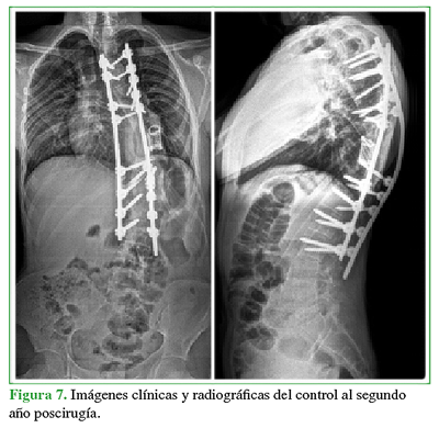
Dystrophic scoliosis in neurofibromatosis is identifiable by being an acute-angle kyphoscoliosis involving a short segment of the spine and producing severe deformity that associated with the dystrophic changes of the spine result in real surgical challenges.We report the clinical case of a 15-year male with severe dystrophic kyphoscoliosis at the thoracolumbar area, with apex at T9, scoliosis with a Cobb angle of 107 °, and segmental kyphosis of 110.7°. The patient underwent a three-stage surgery, performed through a posterior approach, involving a vertebral column resection (VCR) and titanium mesh replacement, and achieving a kyphosis correction of 56% and a scoliosis correction of 59.8%. The patient experienced no major complications nor sequelae and had a favorable course. The VCR is a powerful and demanding surgical technique that allows for the management of the complex kyphoscoliosis deformity to achieve spinal balance; however, it is not without complications, especially neurological and pulmonary complications, which may be unavoidable. Our patient’s quality of life has improved significantly. Key words: Neurofibromatosis; scoliosis; resection; spineLevel of Evidence: IVDownloads
References
Wang Z, Fu C, Leng J, Qu Z, Xu F, Liu Y. Treatment of dystrophic scoliosis in neurofibromatosis type 1 with onestage posterior pedicle screw technique. Spine J 2015;15(4):587.85. https://doi.org/10.1016/j.spinee.2014.10.014
Bersusky E. Deformidades vertebrales por neurofibromatosis. Rev Asoc Argent Ortop Traumatol 1999;64(4):263-9. https://www.aaot.org.ar/revista/1993_2002/1999/1999_4/640401.pdf
Coca Pérez A, Heredero JJ, Burgos J, Ferrero A, Aparicio-Meix JM. Neurofibromatosis tipo 1: escoliosis distrófica y tumor paravertebral. Rev Esp Pediatr 2004;60(3):243-5. https://pesquisa.bvsalud.org/portal/resource/pt/ibc-37743
Woodrow C, Clarke A, Amirfey R. Neurofibromatosis. Orthop Trauma 2015;29(3):206-10.
https://doi.org/10.1016/j.mporth.2015.02.004
Patel N B, Stacy GS. Musculoskeletal manifestations of neurofibromatosis type 1. AJR Am J Roentgenol
;199(1):W99-106. https://doi.org/10.2214/AJR.11.7811
Crawford AH, Herrera-Soto J. Scoliosis associated with neurofibromatosis. Orthop Clin North Am 2007;38(4):553-62. https://doi.org/10.1016/j.ocl.2007.03.008
Haher TR, Gorup JM, Shin TM, Homel P, Merola AA, Grogan DP, et al. Results of the Scoliosis Research Society
instrument for evaluation of surgical outcome in adolescent idiopathic scoliosis. Spine 1999;24(14):1435-40.
https://doi.org/10.1097/00007632-199907150-00008
Boachie-Adjei O, Yagi M, Sacramento-Dominguez C, Akoto H, Cunningham ME, Gupta M, et al. Surgical
risk stratification based on preoperative risk factors in severe pediatric spinal deformity surgery. Spine Deform
;2(5):340-9. https://doi.org/10.1016/j.jspd.2014.05.004
Tejeda Barreras M. Esteroides en lesión medular postraumática aguda. Columna 2011;1(2):39-42.
https://www.medigraphic.com/pdfs/columna/col-2011/col112d.pdf
Motono N, Kawaguchi M, Kawaharab N, Uramoto H. Case report of surgical treatment of scoliosis caused by
neurofibroma located posterior mediastinum. Int J Surg Case Rep 2018;53:168-70.
https://doi.org/10.1016/j.ijscr.2018.10.071
Tsirikos AI, Saifuddin A, Noordeen MH. Spinal deformity in neurofibromatosis type-1: diagnosis and treatment. Eur Spine J 2005;14(5):427-39. https://doi.org/10.1007/s00586-004-0829-7
Larson AN, Ledonio CGT, Brearley AM, Sucato DJ, Carreon LY, Crawford AH, et al. Predictive value and
interrater reliability of radiographic factors in neurofibromatosis patients with dystrophic scoliosis. Spine Deform
;6(5):560-7. https://doi.org/10.1016/j.jspd.2018.02.011
Lenke LG, Newton PO, Sucato DJ, Shufflebarger HL, Emans JB, Sponseller PD, et al. Complications after
consecutive vertebral column resections for severe pediatric spinal deformity. Spine (Phila Pa 1976)
;38(2):119-32. https://doi.org/10.1097/BRS.0b013e318269fab1
Schwab F, Blondel B, Chay E, Demakakos J, Lenke L, Tropiano P, et al. The comprehensive anatomical spinal
osteotomy classification. Neurosurgery 2014;74(1):112-20. https://doi.org/10.1227/01.neu.0000462076.73701.09
Uribe JS, Schwab F, Mundis GM, Xu DS, Januszewski J, Kanter AS, et al. The comprehensive anatomical spinal
osteotomy and anterior column realignment classification. J Neurosurg Spine 2018;29(5):565-75.
https://doi.org/10.3171/2018.4.SPINE171206
Atici Y, Balioglu MB, Kargin D, Mert M, Albayrak A, Kaygusuz MA. Analysis of complications following posterior
vertebral column resection for the treatment of severe angular kyphosis greater than 100°. Acta Orthop Traumatol Turc 2017;51(3):201-8. https://doi.org/10.1016/j.aott.2017.02.015
Auerbach JD, Lenke LG, Bridwell KH, Sehn JK, Milby AH, Bumpass D, et al. Major complications and
comparison between 3-column osteotomy techniques in 105 consecutive spinal deformity procedures. Spine (Phila Pa1976) 2012;37(14):1198-210. https://doi.org/10.1097/BRS.0b013e31824fffde
Wang S, Aikenmu K, Zhang J, Qiu G, Guo J, Zhang Y, Weng, X. The aim of this retrospective study is to evaluate
the efficacy and safety of posterior-only vertebral column resection (PVCR) for the treatment of angular and
isolated congenital kyphosis. Eur Spine J 2017;26(7):1817-25. https://doi.org/10.1007/s00586-015-4344-9
Sacramento-Domínguez C, Yagi M, Ayamga J, Nemani VM, Akoto H, Mahmud R, et al. Apex of deformity for
three column osteotomy. Does it matter in the occurrence of complications? FOCOS Spine Research Group. Spine J 2015;15(11):2351-9. https://doi.org/10.1016/j.spinee.2015.07.010
Sheha ED, Kim HJ, Cunningham ME. Vertebral column resection for complex spinal deformity. Semin Spine Surg 2017;29(4):175-83. https://doi.org/10.1053/j.semss.2017.08.002
Wang H, Guo J, Wang S, Yang Y, Zhang Y, Qiu G, Zhang J. Instrumentation failure after posterior vertebral column resection in adult spinal deformity. Spine (Phila Pa 1976) 2017;42(7):471-8.
https://doi.org/10.1097/BRS.0000000000001844
Sravisht Iyer, Venu M. Nemani, Han Jo Kim. A review of complications and outcomes following vertebral column resection in adults. Asian Spine J 2016;10(3):601-9. https://doi.org/10.4184/asj.2016.10.3.601
Climent JM, Bagó J, Rodríguez-Ruiz C, Sánchez-Raya J, Mulet S, Cholbi F. Nueva estrategia para mejorar la
medida de la calidad de vida en la escoliosis idiopática: adición de la dimensión de deformidad percibida al
cuestionario de la Scoliosis Research Society (SRS-22). Rehabilitación 2011;45(3):228-32.
https://doi.org/10.1016/j.rh.2011.04.007
Merola AA, Haher TR, Brkaric M, Panagopoulos G, Mathur S, Kohani O, et al. A multicenter study of the outcomes of the surgical treatment of adolescent idiopathic scoliosis using the Scoliosis Research Society (SRS) outcome instrument. Spine (Phila Pa 1976) 2002;27(18):2046-51. https://doi.org/10.1097/00007632-200209150-00015
Manuscript acceptance by the Journal implies the simultaneous non-submission to any other journal or publishing house. The RAAOT is under the Licencia Creative Commnos Atribución-NoComercial-Compartir Obras Derivadas Igual 4.0 Internacional (CC-BY-NC.SA 4.0) (http://creativecommons.org/licences/by-nc-sa/4.0/deed.es). Articles can be shared, copied, distributed, modified, altered, transformed into a derivative work, executed and publicly communicated, provided a) the authors and the original publication (Journal, Publisher and URL) are mentioned, b) they are not used for commercial purposes, c) the same terms of the license are maintained.
In the event that the manuscript is approved for its next publication, the authors retain the copyright and will assign to the journal the rights of publication, edition, reproduction, distribution, exhibition and communication at a national and international level in the different databases. data, repositories and portals.
It is hereby stated that the mentioned manuscript has not been published and that it is not being printed in any other national or foreign journal.
The authors hereby accept the necessary modifications, suggested by the reviewers, in order to adapt the manuscript to the style and publication rules of this Journal.


