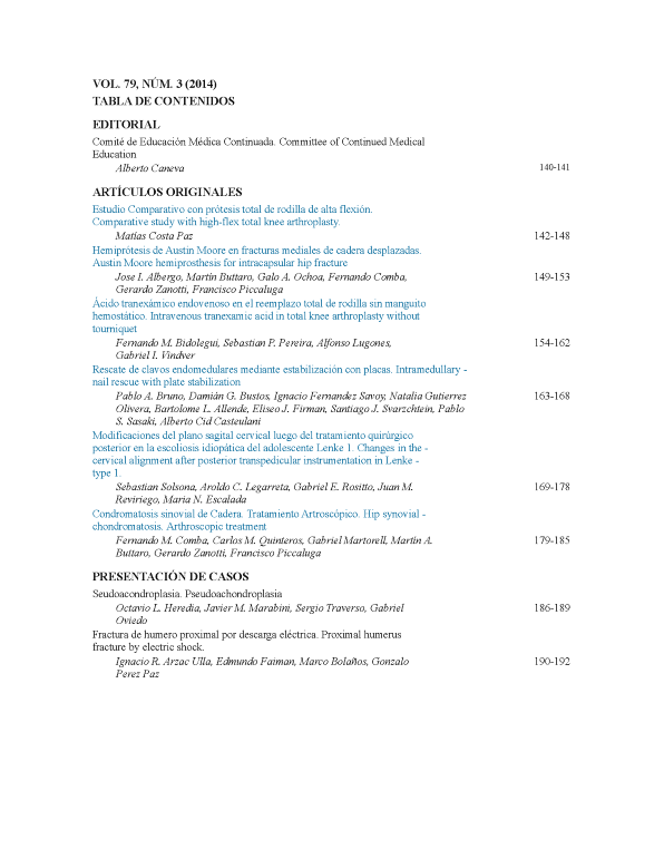
Modificaciones del plano sagital cervical luego del tratamiento quirúrgico posterior en la escoliosis idiopática del adolescente Lenke 1. [Changes in the cervical alignment after posterior transpedicular instrumentation in Lenke type 1 adolescent idiopathic scoliosis].
Contenido principal del artículo
Resumen
Descargas
Métricas
Detalles del artículo

Esta obra está bajo licencia internacional Creative Commons Reconocimiento-NoComercial-CompartirIgual 4.0.
La aceptación del manuscrito por parte de la revista implica la no presentación simultánea a otras revistas u órganos editoriales. La RAAOT se encuentra bajo la licencia Creative Commons 4.0. Atribución-NoComercial-CompartirIgual (http://creativecommons.org/licenses/by-nc-sa/4.0/deed.es). Se puede compartir, copiar, distribuir, alterar, transformar, generar una obra derivada, ejecutar y comunicar públicamente la obra, siempre que: a) se cite la autoría y la fuente original de su publicación (revista, editorial y URL de la obra); b) no se usen para fines comerciales; c) se mantengan los mismos términos de la licencia.
En caso de que el manuscrito sea aprobado para su próxima publicación, los autores conservan los derechos de autor y cederán a la revista los derechos de la publicación, edición, reproducción, distribución, exhibición y comunicación a nivel nacional e internacional en las diferentes bases de datos, repositorios y portales.
Se deja constancia que el referido artículo es inédito y que no está en espera de impresión en alguna otra publicación nacional o extranjera.
Por la presente, acepta/n las modificaciones que sean necesarias, sugeridas en la revisión por los pares (referato), para adaptar el trabajo al estilo y modalidad de publicación de la Revista.
Citas
cervical spine in compression is increased under a follower load. Spine 2000;25:1548-54.
2. Panjabi MM. Cervical spine models for biomechanical research. Spine 1998;23:2684-700.
3. Harrison DD, Harrison DE, Janik TJ, Cailliet R, Ferrantelli JR, Holland B, et al. Modeling of the sagittal cervical spine as a
method of discriminate hypolordosis. Results of elliptical and circular modeling in 72 asymptomatic subjects, 52 acute neck pain
and 70 chronic neck pain subjects. Spine 2004;29(22):2485-92.
4. Reilly CW, Mulpuri K, Perdios A. Evidence-based medicine analysis of all pedicle screw constructs in AIS. Spine
2007;32(19S):109-14.
5. Kim YJ, Lenke LG, Kim J, Bridwell KH, Cho SK, Chen G, et al. Comparative analysis of pedicle screw vs. hybrid
instrumentation in posterior spinal fusion of AIS. Spine 2006;31:291-8.
6. Watanabe K, Lenke LG, Bridwell KH, Kim YJ, Watanabe K, Kim YW, et al. Comparison of radiographic outcomes for the
treatment of scoliotic curves greater than 100 degrees: wires vs. hooks vs. screws. Spine 2008;33:1084-92.
7. Suk SI, Kim WJ, Kim JH, Lee SM. Restoration of thoracic kyphosis in the hypokyphotic spine: a comparison between
multiple-hook and segmental pedicle screw fixation in AIS. J Spinal Disord 1999;12:489-95.
8. Suk SI, Lee SM, Chung ER, Kim JH, Kim WJ, Sohn HM. Determination of distal segmental pedicle screw fixation in single
thoracic idiopathic scoliosis. Spine 2003;28:484-91.
9. Lowenstein JE, Matsumoto H, Vitale MG, Weidenbaum M, Gomez JA, Young-In Lee FYI, et al. Coronal and sagittal plane
correction in AIS: comparison between all pedicle screws vs. hybrid thoracic hook lumbar screws constructs. Spine 2007;32:448-
52.
10. Sucato DJ, Agrawal S, O´Brien MF, Lowe TG, Richards SB, Lenke L. Restoration of thoracic kyphosis after operative
treatment of AIS. A multicenter comparison of three surgical approaches. Spine 2008;30(24):2630-6.
11. Harms Study Group. Preservation of thoracic kyphosis is critical to maintain lumbar lordosis in the surgical treatment of AIS.
Spine 2010;35(14):1365-70.
12. Lee SM, Suk SI, Chung ER. Direct vertebral rotation: a new technique of three-dimensional deformity correction with
segmental pedicle screw fixation in AIS. Spine 2004;29:343-9.
13. Gerald M, Gibson MJ, Quan Y. Correction of main thoracic adolescent idiopathic scoliosis using pedicle screws
instrumentation. Does higher implant density improve correction? Spine 2010;35:562-7.
14. Hillibrand AS, Tannenbaum DA, Graziano GP, Loder RT, Hensinger RN. The sagittal aligment of the cervical spine in
adolescent idiopathic scoliosis. J Pediatr Orthop 1995;15:627-32.
15. Clement JL, Chau E, Kimkpe C, Vallade MJ. Restoration of thoracic kyphosis by posterior instrumentation in AIS.
Comparative radiographic analysis of two methods of reduction. Spine 2008;33(14):1579-87.
16. Westrick ER, Ward TW. AIS: 5-year to 20-year evidence-based surgical results. J Pediatr Orthop 2011;31:s61-68.
17. Harms Study Group. Evaluation of proximal junctional kyphosis in AIS following pedicle screw, hook, or hybrid
instrumentation. Spine 2010;35(2):177-81.
18. Edgar MA, Metha MH. Long term follow up of fused and unfused idiophatic scoliosis. J Bone Joint Surg Br 1988;70:712-6.
19. Moskowitz A, Moe JH, Winter RB, Binner H. Long term follow up of scoliosis fusion. J Bone Joint Surg Am 1980;62:364-76.
20. Canavese F, Turcot K, De Rosa V, de Coulon G, Kaelin A. Cervical spine sagittal aligment variations following posterior spinal
fusion and instrumentation for AIS. Eur Spine J 2011;20:1141-8.

