Kienböck's disease: current concepts
Abstract
Osteonecrosis of the lunate or Kienböck’s disease is a progressive, debilitating condition that can lead to chronic pain and function loss. Despite it was identification almost 100 years ago, the etiology remains unknown, although mechanical, vascular, and traumatic factors have been associated. The natural history of Kienböck’s disease is poorly defined, and the radiographic findings do not always correlate with the clinical findings. There has been some progress in identifying and understanding the progression of the avascular process and its deleterious effects on wrist mechanics. Initial treatment is conservative. Improvement in surgical techniques with vascularized bone grafts from the distal radius may lead to an outcome improvement for patients in the early stages of disease. However, more research is still needed to determine whether this surgical treatment represents an improvement over conventional treatment alternatives. Recent reports of long-term outcomes for radial shortening osteotomy in osteonecrosis early stage patients and for proximal row carpectomy (PRC) in advanced Kienböck’s disease patients reveal that these procedures provide reliable options for the long-term management of this challenging condition.Downloads
References
Wagner JP, Chung KC. A historical report on Robert Kienböck (1871-1953) and Kienböck’s disease. J Hand Surg
Am 2005;30:1117-21. https://doi.org/10.1016/j.jhsa.2005.08.002
Hultén O. Uber anatomische variationen der handgelenkknochen. Acta Radiol Scand 1928;9:155-68.
https://doi.org/10.3109/00016922809176658
Nakamura R, Tsuge S, Watanabe K, Tsunoda K. Radial wedge osteotomy for Kienböck’s disease. J Bone Joint Surg Am 1991;73:1391-6. PMID: 1918122
Gelberman RH, Bauman TD, Menon J, Akeson WH. The vascularity of the lunate bone and Kienböck’s disease.
J Hand Surg Am 1980;5(3):272-8. https://doi.org/10.1016/s0363-5023(80)80013-x
Panagis JS, Gelberman RH, Taleisnik J, Baumgaertner M. The arterial anatomy of the human carpus. Part II: the
intraosseous vascularity. J Hand Surg Am 1983;8(4):375-82. https://doi.org/10.1016/s0363-5023(83)80195-6
Allan CH, Joshi A, Lichtman DM. Kienbock’s disease: diagnosis and treatment. J Am Acad Orthop Surg
;9:128-36. https://doi.org/10.5435/00124635-200103000-00006
Pichler M, Putz R. The venous drainage of the lunate bone. Surg Radiol Anat 2003;24:372-6.
https://doi.org/10.1007/s00276-002-0065-y
Luo J, Diao E. Kienböck’s disease: an approach to treatment. Hand Clin 2006;22:465-73.
https://doi.org/10.1016/j.hcl.2006.07.003
Schuind F, Eslami S, Ledoux P. Kienböck’s disease. J Bone Joint Surg Br 2008;90:133-9.
https://doi.org/10.1302/0301-620X.90B2.20112
Bain GI, Begg M. Arthroscopic assessment and classification of Kienbock’s disease. Tech Hand Up Extrem Surg 2006;10:8-13. https://doi.org/10.1097/00130911-200603000-00003
Goldfarb CA, Hsu J, Gelberman RH, Boyer MI. The Lichtman classification for Kienböck’s disease: an assessment of reliability. J Hand Surg Am 2003;28:74-80. https://doi.org/ https://doi.org/10.1053/jhsu.2003.50035
Salmon J, Stanley JK, Trail IA. Kienböck’s disease: conservative management versus radial shortening. J Bone Joint Surg Br 2000;82:820-3. https://doi.org/10.1302/0301-620x.82b6.10570
Beckenbaugh RD, Shives TC, Dobyns JH, Linscheid RL. Kienböck’s disease: the natural history of Kienböck’s
disease and considerations of lunate fractures. Clin Orthop 1980;149:98-106. PMID: 7408323
Keith PP, Nuttall D, Trail I. Long-term outcome of nonsurgically managed Kienböck’s disease. J Hand Surg Am
;29:63-7. https://doi.org/10.1016/j.jhsa.2003.10.016
Delaere O, Dury M, Molderez A, Foucher G. Conservative versus operative treatment for Kienböck’s disease: a
retrospective study. J Hand Surg Br 1998;23:33-6. https://doi.org/10.1016/s0266-7681(98)80214-3
Lichtman DM, Roure AR. External fixation for the treatment of Kienböck’s disease. Hand Clin 1993;9:691-7.
PMID: 8300738
Moran SL, Shin AY. Vascularized bone grafting for the treatment of carpal pathology. Orthop Clin North Am
;38:73-85. https://doi.org/10.1016/j.ocl.2006.10.010
Daecke W, Lorenz S, Wieloch P, Jung M, Martini AK. Vascularized os pisiform for reinforcement of the lunate in
Kienböck’s disease: an average of 12 years of follow-up study. J Hand Surg Am 2005;30:915-22.
https://doi.org/10.1016/j.jhsa.2005.03.019
Sheetz KK, Bishop AT, Berger RA. The arterial blood supply of the distal radius and ulna and its potential use in vascularized pedicled bone grafts. J Hand Surg Am 1995;20:902-14. https://doi.org/10.1016/S0363-5023(05)80136-4
Bochud RC, Büchler U. Kienböck’s disease, early stage 3: height reconstruction and core revascularization of the lunate. J Hand Surg Br 1994;19:466-78. https://doi.org/10.1016/0266-7681(94)90212-7
Tamai S, Yajima H, Ono H. Revascularization procedures in the treatment of Kienböck’s disease. Hand Clin
;9:455-66. PMID: 8408255
Moran SL, Cooney WP, Berger RA, Bishop AT, Shin AY. The use of the 4-5 extensor compartmental vascularized bone graft for the treatment of Kienbock’s disease. J Hand Surg Am 2005;30:50-8.
https://doi.org/10.1016/j.jhsa.2004.10.002
Gabl M, Lutz M, Reinhart C, Zimmerman R, Pechlaner S, Hussl H, et al. Stage 3 Kienböck’s disease: reconstruction of the fractured lunate using a free vascularized iliac bone graft and external fixation. J Hand Surg Br 2002;27:369-73. https://doi.org/10.1054/jhsb.2002.0766
Arora R, Lutz M, Deml C, Krappinger D, Zimmermann R, Gabl M. Long-term subjective and radiological outcome after reconstruction of Kienböck’s disease stage 3 treated by a free vascularized iliac bone graft. J Hand Surg Am 2008;33:175-81. https://doi.org/10.1016/j.jhsa.2007.11.005
Raven EE, Haverkamp D, Marti RK. Outcome of Kienböck’s disease 22 years after distal radius shortening
osteotomy. Clin Orthop Relat Res 2007;460:137-41. https://doi.org/10.1097/BLO.0b013e318041d309
Watanabe T, Takahara M, Tsuchida H, Yamahara S, Kikuchi N, Ogino T. Long-term follow-up of radial shortening osteotomy for Kienböck disease. J Bone Joint Surg Am 2008;90(8):1705-11. https://doi.org/10.2106/JBJS.G.00421
Altay T, Kaya A, Karapinar L, Ozturk H, Kayali C. Is radial shortening useful for Litchman stage 3B Kienbock’s
disease? Int Orthop 2008;32(6):747-52. https://doi.org/10.1007/s00264-007-0428-4
Watanabe K, Nakamura R, Horii E, Miura T. Biomechanical analysis of radial wedge osteotomy for the treatment of Kienböck’s disease. J Hand Surg Am 1993;18:686-90. https://doi.org/10.1016/0363-5023(93)90319-X
Koh S, Nakamura R, Horii E, Nakao E, Inagaki H, Yajima H. Surgical outcome of radial osteotomy for Kienböck’s disease—minimum 10 years of follow-up. J Hand Surg Am 2003;28:910-6. https://doi.org/10.1016/s0363-5023(03)00490-8
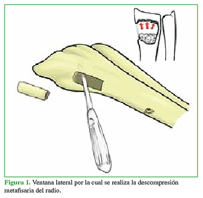
Illarramendi AA, Schulz C, De Carli P. The surgical treatment of Kienböck’s disease by radius and ulna metaphyseal core decompression. J Hand Surg Am 2001;26:252-60. https://doi.org/10.1053/jhsu.2001.22928
De Carli P, Zaidenberg EE, Alfie V, Donndorff A, Boretto JG, Gallucci GL. Radius core decompression for
Kienböck disease stage IIIA: outcomes at 13 years follow-up. J Hand Surg Am 2017;42(9):752.e1-752.e6.
https://doi.org/10.1016/j.jhsa.2017.05.017
Van den Dungen S, Dury M, Foucher G, Marin Braun F, Loréa P. Conservative treatment versus
scaphotrapeziotrapezoid arthrodesis for Kienböck’s disease. A retrospective study. Chir Main 2006;25:141-5.
https://doi.org/10.1016/j.main.2006.07.030
Tambe AD, Trail IA, Stanley JK. Wrist fusion versus limited carpal fusion in advanced Kienbock’s disease. Int
Orthop 2005;29:355-8. https://doi.org/10.1007/s00264-005-0013-7
Salomon GD, Eaton RG. Proximal row carpectomy with partial capitate resection. J Hand Surg Am 1996;21:2-8.
https://doi.org/10.1016/S0363-5023(96)80147-X
Croog AS, Stern PJ. Proximal row carpectomy for advanced Kienböck’s disease: average 10-year follow-up. J Hand Surg Am 2008;33:1122-30. https://doi.org/10.1016/j.jhsa.2008.02.031
DiDonna ML, Kiefhaber TR, Stern PJ. Proximal row carpectomy: study with a minimum of ten years of follow-up. J Bone Joint Surg Am 2004;86:2359-65. PMID: 15523004
De Smet L, Robijns P, Degreef I. Proximal row carpectomy in advanced Kienböck’s disease. J Hand Surg Br
;30:585-7. https://doi.org/10.1016/j.jhsb.2005.06.024
Lumsden BC, Stone A, Engber WD. Treatment of advanced-stage Kienböck’s disease with proximal row
carpectomy: an average 15- year follow-up. J Hand Surg Am 2008;33:493-502. https://doi.org/10.1016/j.jhsa.2007.12.010
Schweizer A, von Känel O, Kammer E, Meuli-Simmen C. Long-term follow-up evaluation of denervation of the
wrist. J Hand Surg Am 2006;31:559-64. https://doi.org/10.1016/j.jhsa.2005.12.012
Manuscript acceptance by the Journal implies the simultaneous non-submission to any other journal or publishing house. The RAAOT is under the Licencia Creative Commnos Atribución-NoComercial-Compartir Obras Derivadas Igual 4.0 Internacional (CC-BY-NC.SA 4.0) (http://creativecommons.org/licences/by-nc-sa/4.0/deed.es). Articles can be shared, copied, distributed, modified, altered, transformed into a derivative work, executed and publicly communicated, provided a) the authors and the original publication (Journal, Publisher and URL) are mentioned, b) they are not used for commercial purposes, c) the same terms of the license are maintained.
In the event that the manuscript is approved for its next publication, the authors retain the copyright and will assign to the journal the rights of publication, edition, reproduction, distribution, exhibition and communication at a national and international level in the different databases. data, repositories and portals.
It is hereby stated that the mentioned manuscript has not been published and that it is not being printed in any other national or foreign journal.
The authors hereby accept the necessary modifications, suggested by the reviewers, in order to adapt the manuscript to the style and publication rules of this Journal.