
Análisis tomográfico de la alineación del retropié en pacientes con coaliciones tarsianas
Resumen
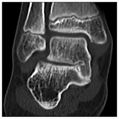
Introducción: El objetivo de este estudio es describir la morfología del retropié mediante cortes coronales con tomografía computarizada en pacientes con coaliciones tarsianas. Materiales y Métodos: Se incluyeron 85 pies de 78 pacientes de entre 9 y 17 años. Fueron divididos en 3 grupos: 1) grupo de control (n = 29), 2) con coaliciones calcáneo-escafoideas (CCE) (n = 31) y 3) con coaliciones astrágalo-calcáneas (CAC) (n = 25). Dos observadores valoraron cinco medidas: Inftal-Suptal, Inftal-Hor, Inftal-Supcal, Suptal-Infcal y el ángulo astrágalo-calcáneo (AAC). Resultados: Los grupos no presentaron diferencias en la distribución por edad y sexo. Los pacientes con coaliciones tarsianas tuvieron valores significativamente superiores en todas las mediciones comparados con el grupo de control (p <0,05 Kruskall-Wallis/ANOVA). Las mediciones del AAC en los pacientes con CCE y CAC fueron significativamente superiores a las del grupo de control (10,09 ± 4,60; 17,77 ± 11,28 y 28,66 ± 8,89, respectivamente, p <0,0001). La distribución del AAC fue muy variable en los pacientes con CCE, mientras que, en la mayoría del grupo CAC, tuvo un patrón de alineación en valgo. No hubo una correlación directa entre los valores del AAC e Inftal-Hor (Spearman 0,27013; p = 0,1916). Conclusiones: En los pacientes con coaliciones tarsianas, la orientación del valgo del retropié suele estar aumentada. La magnitud de esta deformidad es mayor en pacientes con coaliciones CAC, mientras que, en aquellos con CCE pueden manifestarse con una gran variabilidad. El aumento del valgo del retropié no implica necesariamente un aumento de la inclinación de la articulación subastragalina, por lo que esta última debe evaluarse por separado en la planificación preoperatoria. Nivel de Evidencia: IIIDescargas
Citas
Kernbach KJ. Tarsal coalitions: etiology, diagnosis, imaging, and stigmata. Clin Podiatr Med Surg 2010;27(1):105-17. https://doi.org/10.1016/j.cpm.2009.08.006
Cooperman DR, Janke BE, Gilmore A, Latimer BM, Brinker MR, Thompson GH. A three-dimensional study of
calcaneonavicular tarsal coalitions. J Pediatr Orthop 2001;21(5):648-51. PMID: 11521035
Stormont DM, Peterson HA. The relative incidence of tarsal coalition. Clin Orthop 1983;(181):28-36.
PMID: 6641062
Masquijo JJ, Jarvis J. Associated talocalcaneal and calcaneonavicular coalitions in the same foot. J Pediatr Orthop B 2010;19(6):507-10. https://doi.org/10.1097/BPB.0b013e32833ce484
Mubarak SJ, Patel PN, Upasani VV, Moor MA, Wenger DR. Calcaneonavicular coalition: treatment by excision and fat graft. J Pediatr Orthop 2009;29(5):418-26. https://doi.org/10.1097/BPO.0b013e3181aa24c0
Kothari A, Masquijo J. Surgical treatment of tarsal coalitions in children and adolescents. EFORT Open Rev
;5(2):80-9. http://doi.org/10.1302/2058-5241.5.180106
Masquijo JJ, Vazquez I, Allende V, Lanfranchi L, Torres-Gomez A, Dobbs MB. Surgical reconstruction for
talocalcaneal coalitions with severe hindfoot valgus deformity. J Pediatr Orthop 2017;37(4):293-7. https://doi.org/10.1097/BPO.0000000000000642
Probasco W, Haleem AM, Yu J, Sangeorzan BJ, Deland JT, Ellis SJ. Assessment of coronal plane subtalar joint
alignment in peritalar subluxation via weight-bearing multiplanar imaging. Foot Ankle Int 2015;36(3):302-9.
https://doi.org/10.1177/1071100714557861
Masquijo JJ, Torres-Gomez A, Tourn D. Fiabilidad del ángulo astrágalo-calcáneo. Rev Esp Cir Ortop Traumatol
;63(1):20-3. https://doi.org/10.1016/j.recot.2018.08.003
Wilde PH, Torode IP, Dickens DR, Gole WG. Resection for symptomatic talocalcaneal coalition. J Bone Joint Surg Br 1994;76(5):797-801. PMID: 8083272
Upasani VV, Chambers RC, Mubarak SJ. Analysis of calcaneonavicular coalitions using multi-planar threedimensional computed tomography. J Child Orthop 2008;2:301-7. https://doi.org/10.1007/s11832-008-0111-3
Rozansky A, Varley E, Moor M, Wenger DR, Mubarak SJ. A radiologic classification of talocalcaneal coalitions
based on 3D reconstruction. J Child Orthop 2010;4(2):129-35. https://doi.org/10.1007/s11832-009-0224-3
Kemppainen J, Pennock AT, Roocroft JH, Bastrom TP, Mubarak SJ. The use of a portable CT scanner for the
intraoperative assessment of talocalcaneal coalition resections. J Pediatr Orthop 2014;34(5):559-64.
https://doi.org/10.1097/BPO.0000000000000176
Aibinder WR, Young EY, Milbrandt TA. Intraoperative three-dimensional navigation for talocalcaneal coalition
resection. J Foot Ankle Surg 2017;56(5):1091-4. https://doi.org/10.1053/j.jfas.2017.05.046
Stokman JJ, Mitchell J, Noonan K. Subtalar coalition resection utilizing live navigation: a technique tip. J Child
Orthop 2018;12(1):42-6. https://doi.org/10.1302/1863-2548.12.170131
de Wouters S, Tran Duy K, Docquier PL. Patient-specific instruments for surgical resection of painful tarsal
coalition in adolescents. Orthop Traumatol Surg Res 2014;100(4):423-7. https://doi.org/10.1016/j.otsr.2014.02.009
Sobrón FB, Benjumea A, Alonso MB, Parra G, Pérez-Mañanes R, Vaquero J. 3D printing surgical guide for
talocalcaneal coalition resection: technique tip. Foot Ankle Int 2019;40(6):727-32. https://doi.org/10.1177/1071100719833665
Mosca VS, Bevan WP. Talocalcaneal tarsal coalitions and the calcaneal lengthening osteotomy: the role of deformity correction. J Bone Joint Surg Am 2012;94(17):1584-94. https://doi.org/10.2106/JBJS.K.00926
El Shazly O, Mokhtar M, Abdelatif N, Hegazy M, El Hilaly R, El Zohairy A, et al. Coalition resection and medial
displacement calcaneal osteotomy for treatment of symptomatic talocalcaneal coalition: functional and clinical
outcome. Int Orthop 2014;38(12):2513-7. https://doi.org/10.1007/s00264-014-2535
Gantsoudes GD, Roocroft JH, Mubarak SJ. Treatment of talocalcaneal coalitions. J Pediatr Orthop 2012;32(3):301-7. https://doi.org/10.1097/BPO.0b013e318247c76e
Hamel J. Resection of talocalcaneal coalition in children and adolescents without and with osteotomy of the
calcaneus. Oper Orthop Traumatol 2009;21(2):180-92. https://doi.org/10.1007/s00064-009-1706-7
Lisella JM, Bellapianta JM, Manoli A 2nd. Tarsal coalition resection with pes planovalgus hindfoot reconstruction. J Surg Orthop Adv 2011;20(2):102-5. PMID: 21838070
Mahan ST, Spencer SA, Vezeridis PS, Kasser JR. Patient-reported outcomes of tarsal coalitions treated with surgical excision. J Pediatr Orthop 2015;35(6):583-8. https://doi.org/10.1097/BPO.0000000000000334
Quinn EA, Peterson KS, Hyer CF. Calcaneonavicular coalition resection with pes planovalgus reconstruction. J Foot Ankle Surg 2016;55(3):578-82. https://doi.org/10.1053/j.jfas.2016.01.048
Lintz F, de Cesar Netto C, Barg A, Burssens A, Richter M; Weight Bearing CT International Study Group. Weightbearing cone beam CT scans in the foot and ankle. EFORT Open Rev 2018;3(5):278-86.
https://doi.org/10.1302/2058-5241.3.170066
de Cesar Netto C, Schon LC, Thawait GK, Furtado da Fonseca L, Chinanuvathana A, Zbijewski WB, et al. Flexible adult acquired flatfoot deformity: comparison between weight-bearing and non-weight-bearing measurements using cone-beam computed tomography. J Bone Joint Surg Am 2017;99(18):e98. https://doi.org/10.2106/JBJS.16.01366
La aceptación del manuscrito por parte de la revista implica la no presentación simultánea a otras revistas u órganos editoriales. La RAAOT se encuentra bajo la licencia Creative Commons 4.0. Atribución-NoComercial-CompartirIgual (http://creativecommons.org/licenses/by-nc-sa/4.0/deed.es). Se puede compartir, copiar, distribuir, alterar, transformar, generar una obra derivada, ejecutar y comunicar públicamente la obra, siempre que: a) se cite la autoría y la fuente original de su publicación (revista, editorial y URL de la obra); b) no se usen para fines comerciales; c) se mantengan los mismos términos de la licencia.
En caso de que el manuscrito sea aprobado para su próxima publicación, los autores conservan los derechos de autor y cederán a la revista los derechos de la publicación, edición, reproducción, distribución, exhibición y comunicación a nivel nacional e internacional en las diferentes bases de datos, repositorios y portales.
Se deja constancia que el referido artículo es inédito y que no está en espera de impresión en alguna otra publicación nacional o extranjera.
Por la presente, acepta/n las modificaciones que sean necesarias, sugeridas en la revisión por los pares (referato), para adaptar el trabajo al estilo y modalidad de publicación de la Revista.


